SEM Upgrades
Modernize your SEM to the latest technology
Enhance your trusty electron microscope to meet modern standards! Our SEM Upgrade caters to all types of scanning electron microscopes (SEMs), and exchanges outdated or not longer servicable add-ons for cutting-edge technology - from acquisition to control and software. Each microscope configuration and installation is carefully tailored to each customer's requirements.
The point electronic SEM Upgrade adds state of the art performance and keeps the electron microscope serviceable for the next 10+ years. So: Don’t scrap – modernize!
Every modernization is a bespoke solution
We provide you with an upgrade customized to your specific needs. From the required technical capabilities, to the technologies and applications you want to run in the future, to the packaging solution in the plinth or in a separate rack.
We would be happy to advise you.
Advantages
The effects of our modernization for your SEM:
increased performance - from sample insertion to final image
a system that is fully serviceable, incl. 10+ years spare part guarantee for point electronic products
new techniques and capabilities in the SEM
Keep established workflows and add-ons
cost efficient, plus costs savings for acquisition, operation and service
How does the SEM Modernization work?
We optimize your microscope exactly to your needs - but that doesn't mean we exchange the whole system. With our SEM Upgrade, we change everything that needs changing and keep what is important.
YOU KEEP
Your column incl. HV-PSU and pumps
Your add-ons, according to functionality and your requirements
(such as EDX, EBSD, etc).
Your established/certified workflows
Your laboratory layout and ancillary equipment
WE ADD
State of the art electronics
User friendly software with efficient workflows
Improved performance
Network compatibility
New techniques
Reduced costs
Environmental friendlyness
How do we do the SEM Modernization?
Once we have configured the upgrade to your needs, specifications and conditions in your lab, we arrange an appointment at your premises:
- We handle the deinstallation of your old equipment.
- We do the on-site installation of the SEM upgrade for you, in most cases directly in your lab.
- Upgrades take around 3 days. This keeps downtime to a minimum.
- With our included training you will be fit for our applications within 1 day.
Arrange a demo with us or request a non-binding quote.
Our experience
We can look back on around 200 installations of our SEM upgrade. For these microscope models, among others:
- ZEISS / LEO
- PHILIPS / FEI
- Cambridge
- Jeol
- Hitachi
- TESCAN
What is included?
Controls and aquisition
- New SEM control electronics and software
- Support for MicrosoftWindows 7 up to 11, with full network compatibility
- Universal GUI for all upgraded tools independent of manufacturer
- Versatile and fast Scanning system:
- Simultaneous signal acquisition for up to 20 signals (SE, BSE, EDS, WDS, etc.)
- Image resolution of up to 500 MPixels
- Scanning speed down to 10ns dwell time
- Automatic vacuum, gun, stage and detector controls
- New hard panels for column, detector and stage control
- IR chamberscope (for most models)
- PC and display(s) or laptop (optional)
Techniques
- Atomic-number discrimination with BSE detectors
- Topography measurements with shape-from-shadow
- Micro-analysis with EDS detectors and mapping
- Electrical failure analysis with EBIC and EBAC/RCI
New electronics for the complete SEM
- New control of gun HV and electron detectors
- New bipolar or unipolar high power supply
- New scan module for single or double deflection coils
- 12 digital input signals (X-ray mapping)
- 19 inch, 10U form factor size
- Standard USB 2.0 interface
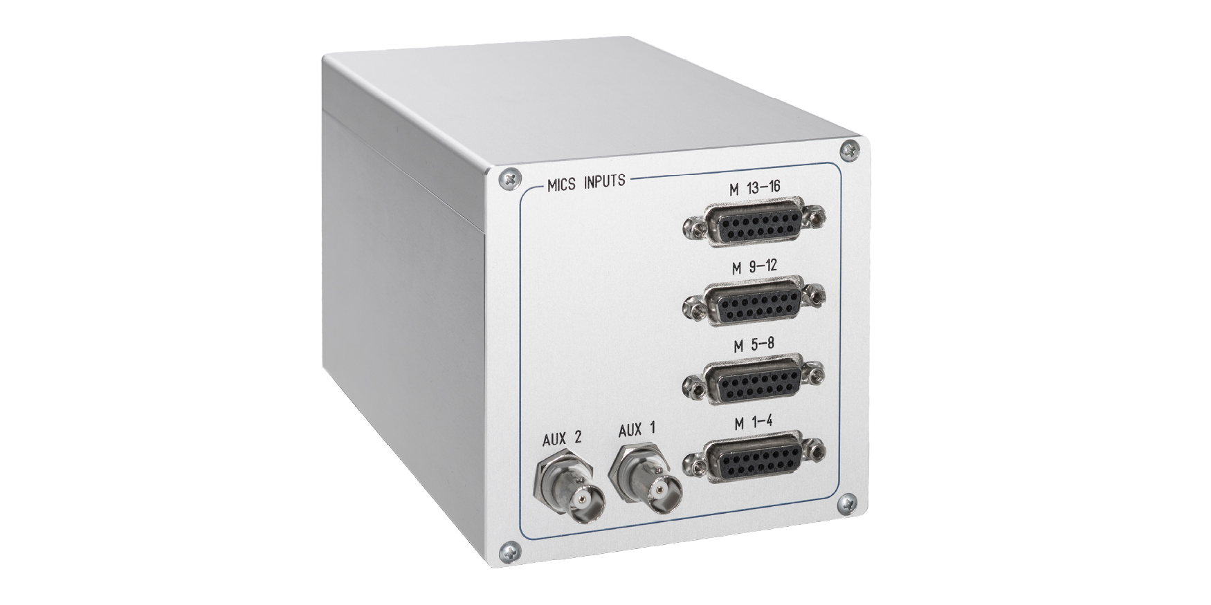
MICS-4 signal amplifier
- For extendend imaging channels, from 4x to 16x
- Channel independendt controls for brightness and contrast
- Advanced input offset and gain controls and calibration
- USB2 controlled and fully integrated with the microscope control software
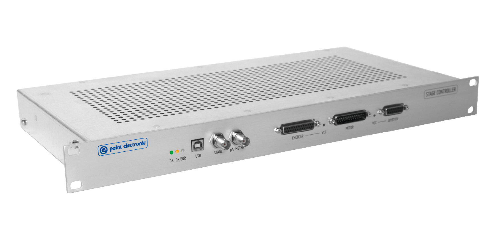
Stage controller
- For automatic positioning and rotation
- Integrated click-and-move, rotation and large area map acquisition
- Configurable software limits for collision avoidance
- USB2 controlled and fully integrated with the microscope control software
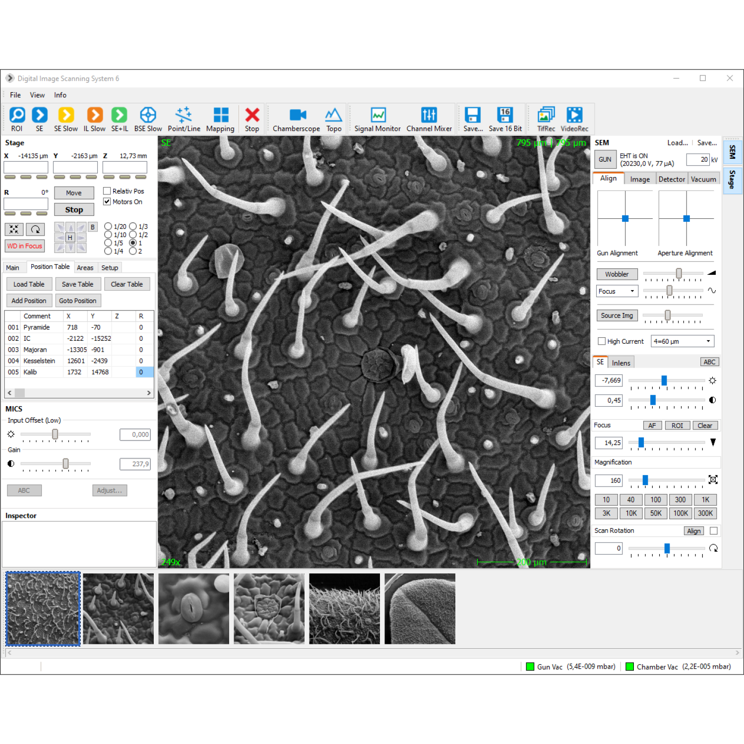
SEM control software
- Control of coils and detectors
- Display of measured values
- Load and save of SEM parameters
- Auto-function for focus/stigmator and brightness/contrast
- TV scan, slow scan, mapping, line scan, point measurements
- Reduced area scan with zoom
- AVI-function with time lapse
- Configurable image acquisition functions for routine actions: Scan buttons can be added, configured and labeled so as signal sources, image resolution, image format
- Integrated vacuum-controller, visualization of vacuum scheme
- Integrated sample stage controller – preset, load and save sample- and stage positions
- Signal monitor with live gradation-curve
- Display and setting of ir-chamberscope
- Integrated image processing software
- Live 3D reconstruction with BSE-detector (optional)
- AutoSEM for automated capture of multiple sample locations (optional)
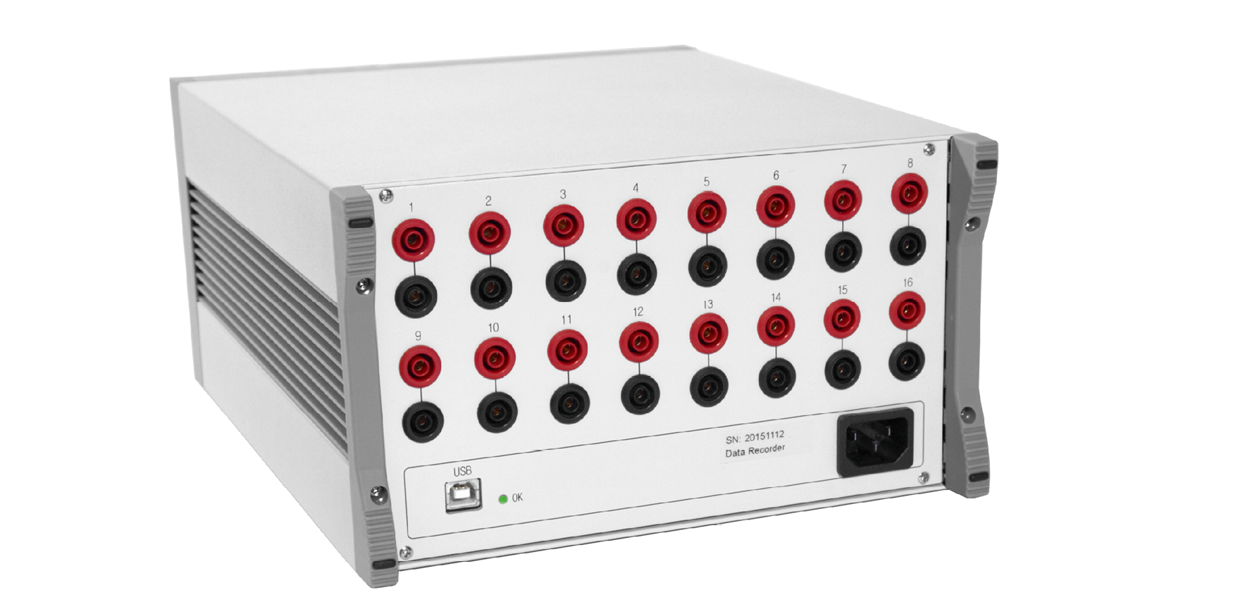
Activity monitor
- For detailed information and preventive maintenance
- Automatic acquisition and complression of microscope operation parameters
- 16x 16-bit analog signal inputs, and software API for loggin events
- Live and offline viewer with PDF exoport
- USB 2.0 controlled and integrated with the new microscope control software
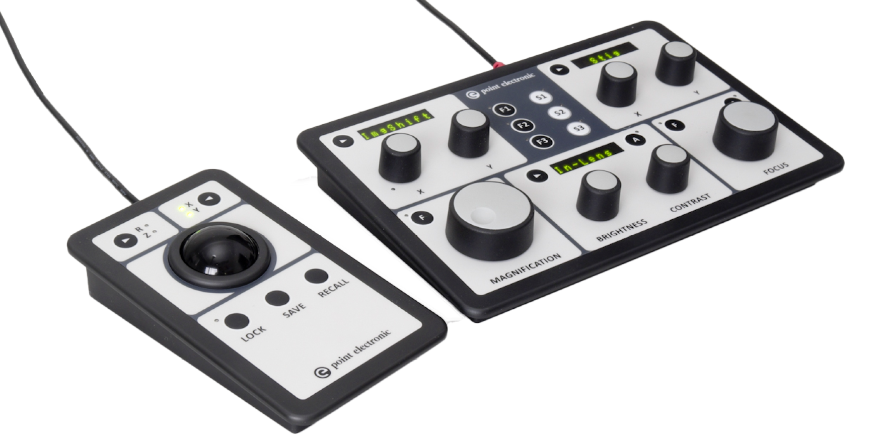
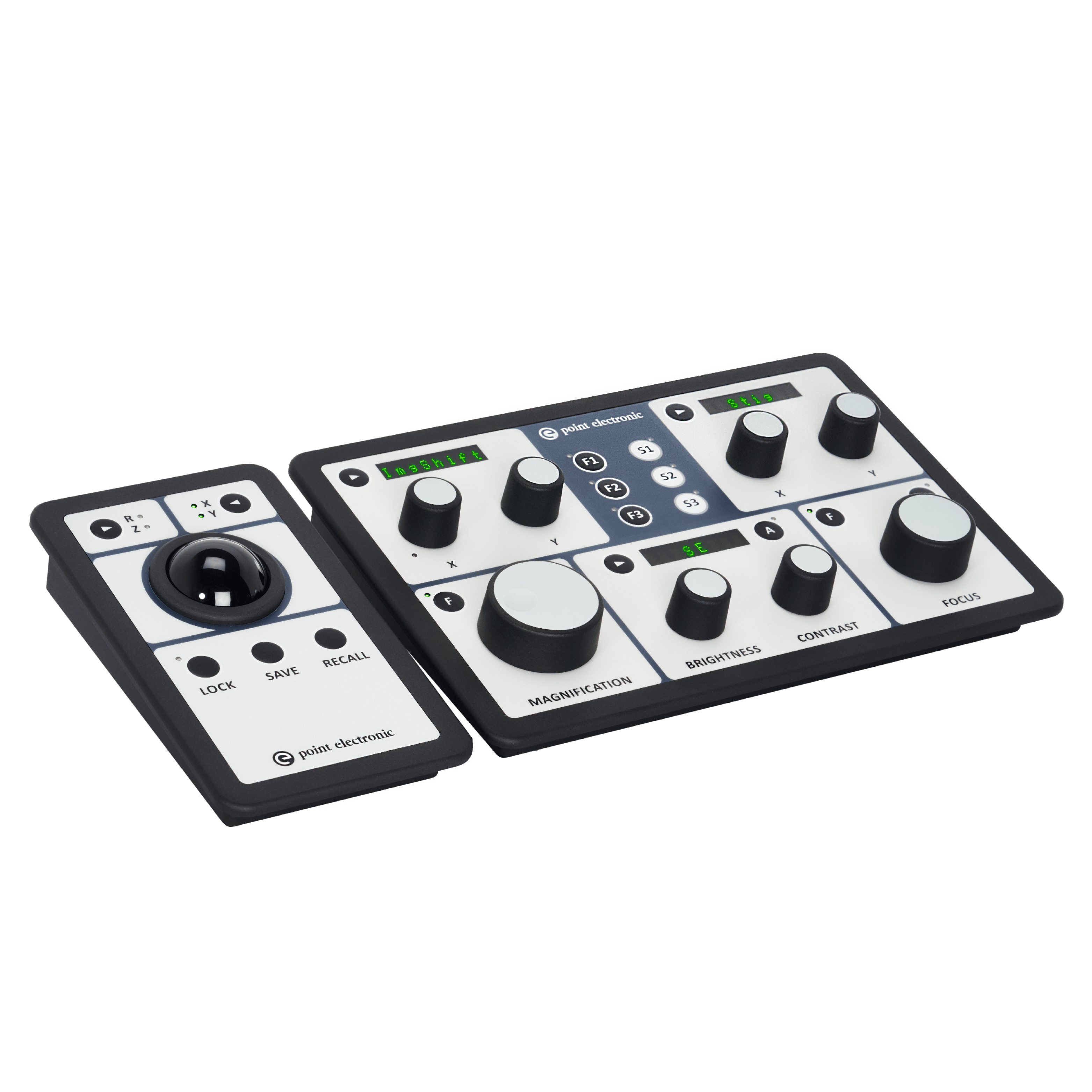
Control panels
- For increased speed and productivity of experienced users
- SEM panel with magnification, focus, image shift, brighness, contrast, stigmatism, etc.
- Stage panel with trackball or joystick, XYRZ locks and store/recall functions
- USB2 controlled and fully integrated wth the microscope control software
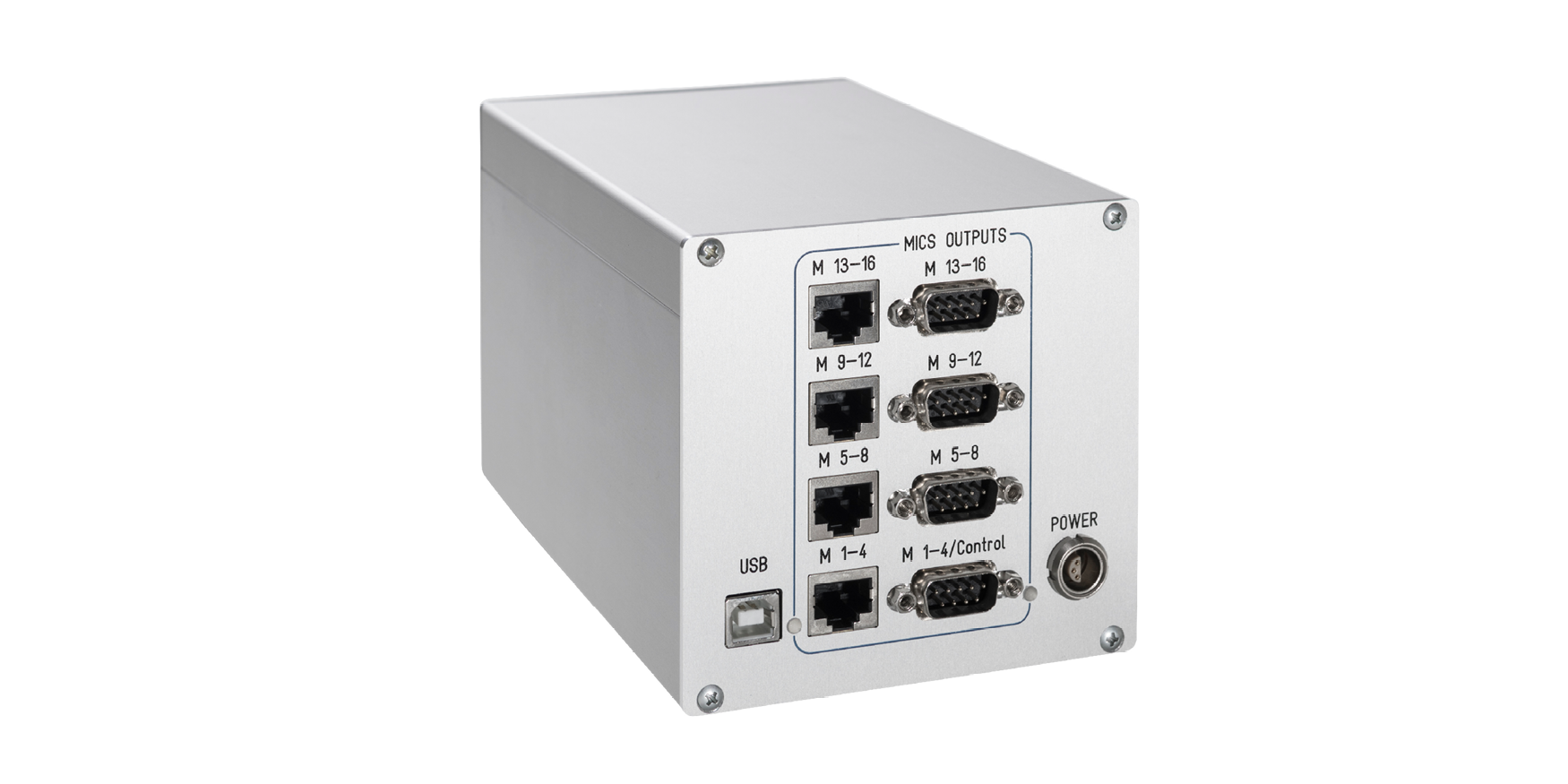
MICS 8 / 12 / 16 signal amplifier
- For extendend imaging channels, by 8x, 12x or 16x
- Channel independendt controls for brightness and contrast
- Advanced input offset and gain controls and calibration
- USB2 controlled and fully integrated with the microscope control software
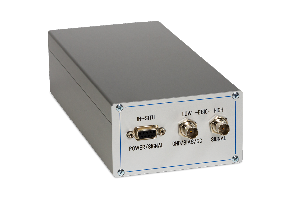
EA amplifiers
- Calibrated high-speed electronics, with in-situ and ex-situ preamplifiers
- Complex multi-stage amplification for imaging, with automatic low-pass filter
- Integrated pico-ammeter for beam current measurements
- Integrated voltage source for biasing
- Integrated current source for compensation
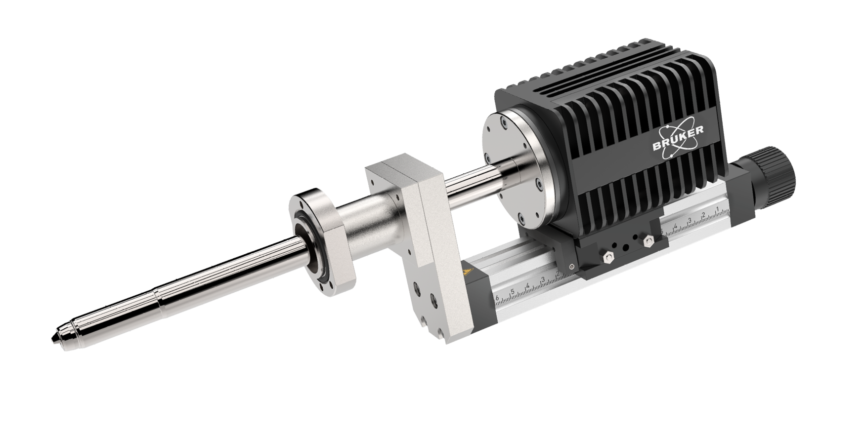
EDS detector
- QUANTAX Compact system by Bruker, containing XFlash® 730M silicon drift detector (SDD), an electronic module, and ESPRIT Compact software
- Allows line scanning and spectral element mapping
- Qualitative and quantitative material analyses
- Fast analysis and reporting
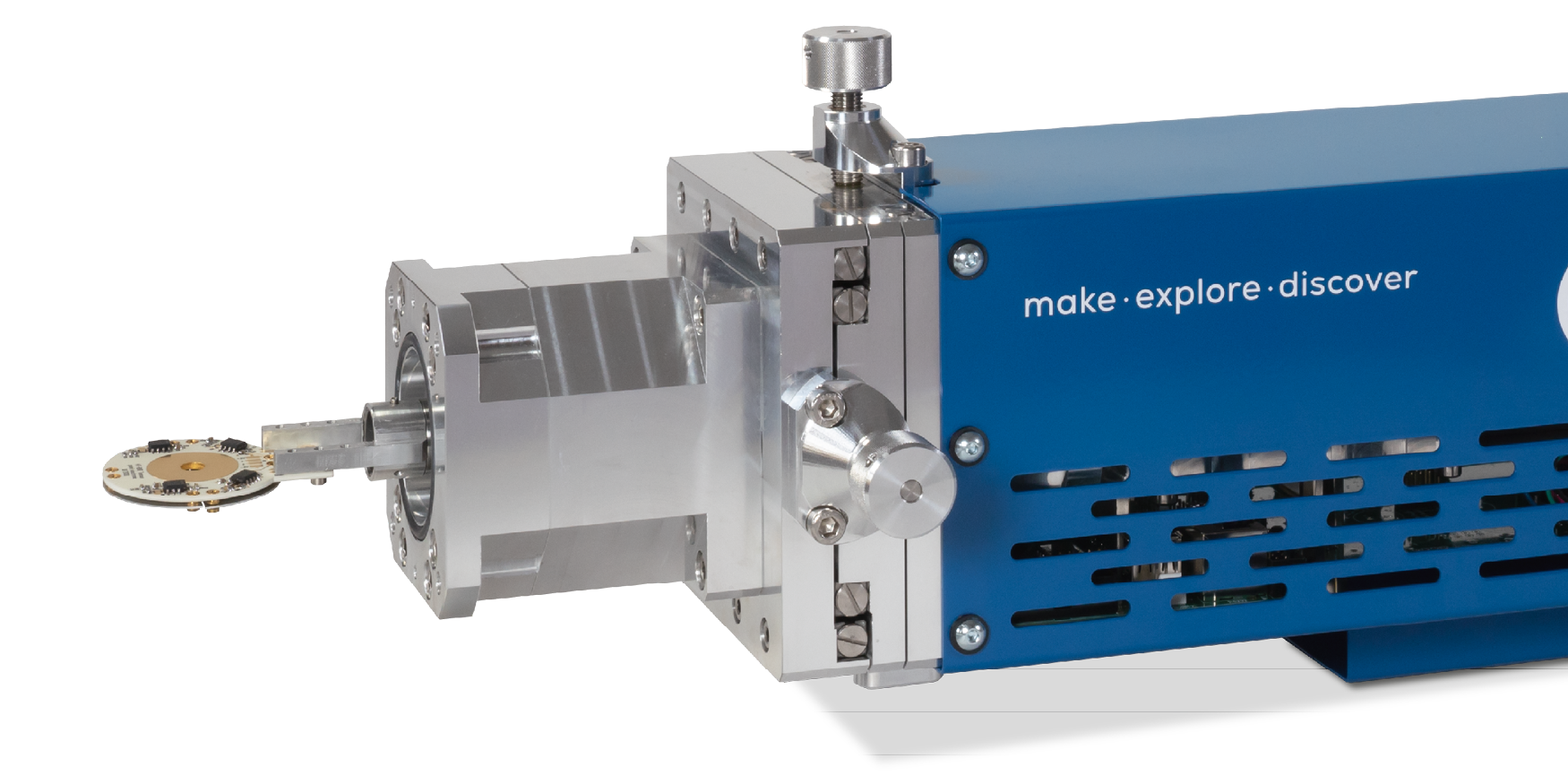
HT BSE detector
- 4Q segmented electrodes with built-in biasing
- Electrodes are light-blind and compatible with laser heating
- Bias voltage to repel secondary electrons and thermal electrons
- Electrodes can be coated in various materials
- easy to disassemble, cleane and recoat
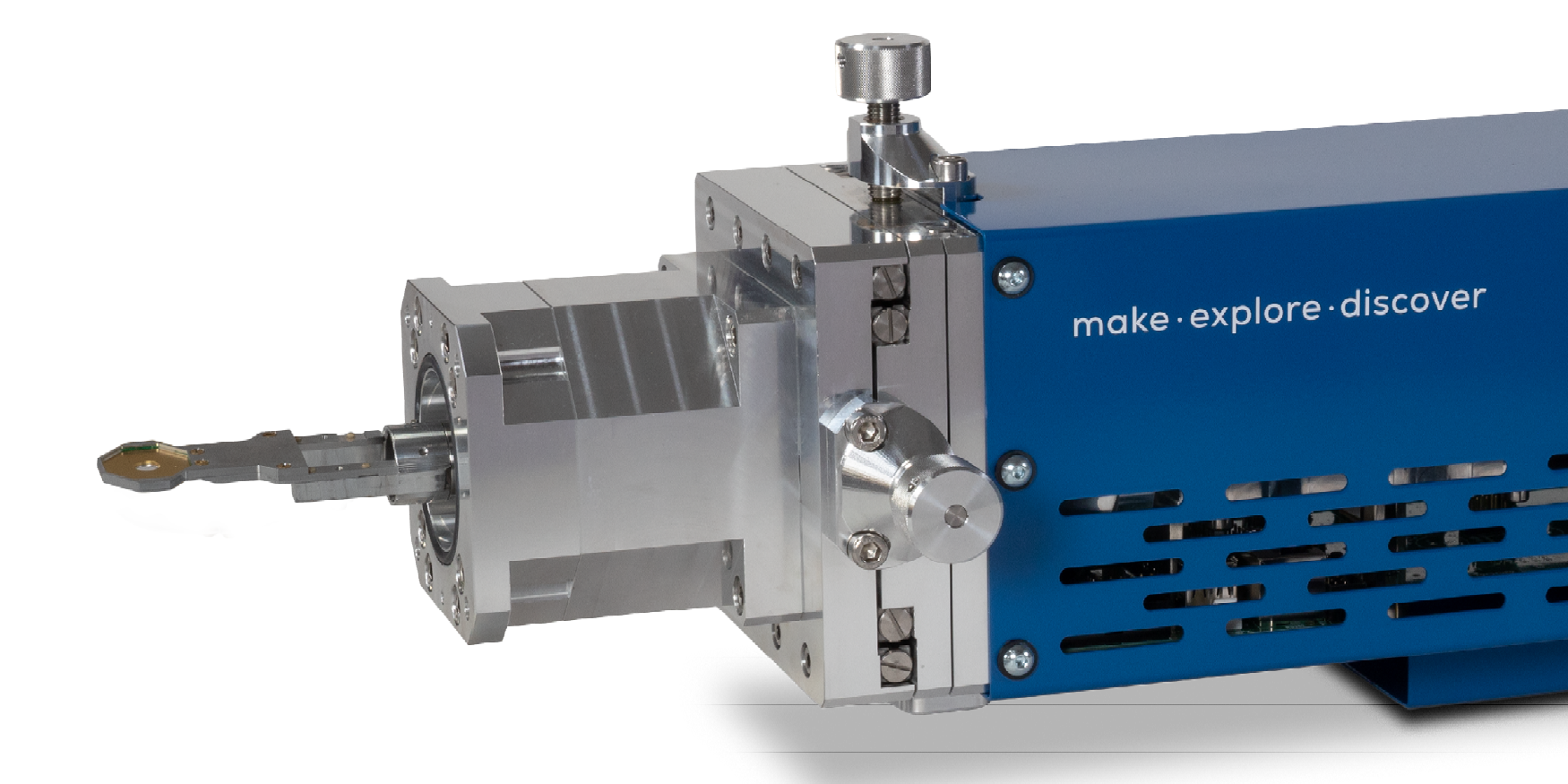
Premium BSE detector
- Four quadrant Si sensor for advanced BSE work
- In-situ preamplification for optimum efficiency and speed
- Optional low-kV sensors
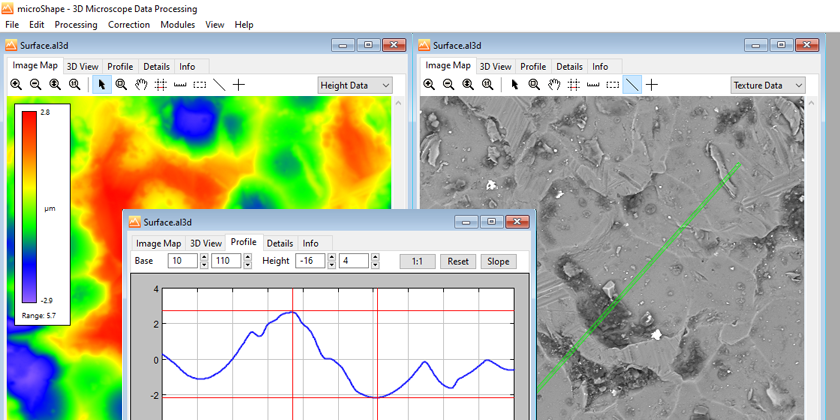
microShape
- Offline 3D viewer of BSE topography data
- Measurement of line profiles, heights, distances and angles in 3D
- 3D data processing including data correction and overlay
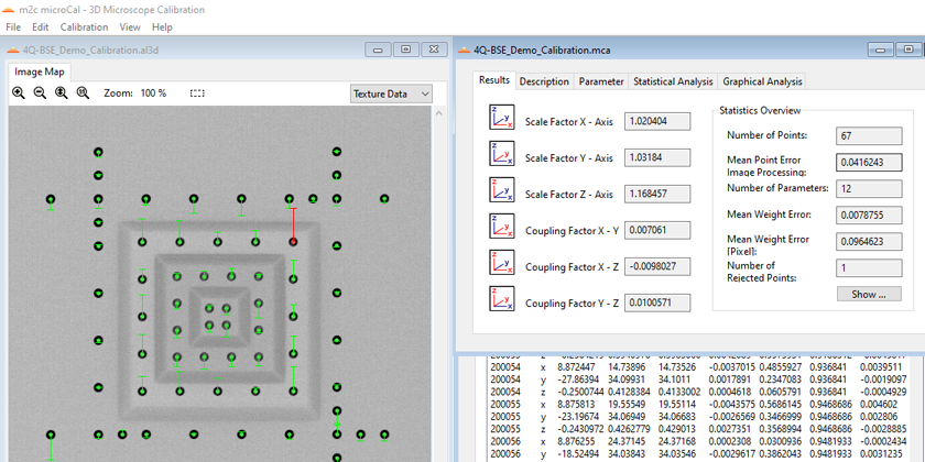
microCal
- Offline 3D calibration and reporting software
- Automated calculation of scales and shearing between all coordinate axes
- Analyses of non-linear scanning deviations
Further options
Additional add-ons and software on demand.
Have a look at our portfolio or contact us.
-
Comparison: SEM before and after the upgrade by point electronic GmbH

-
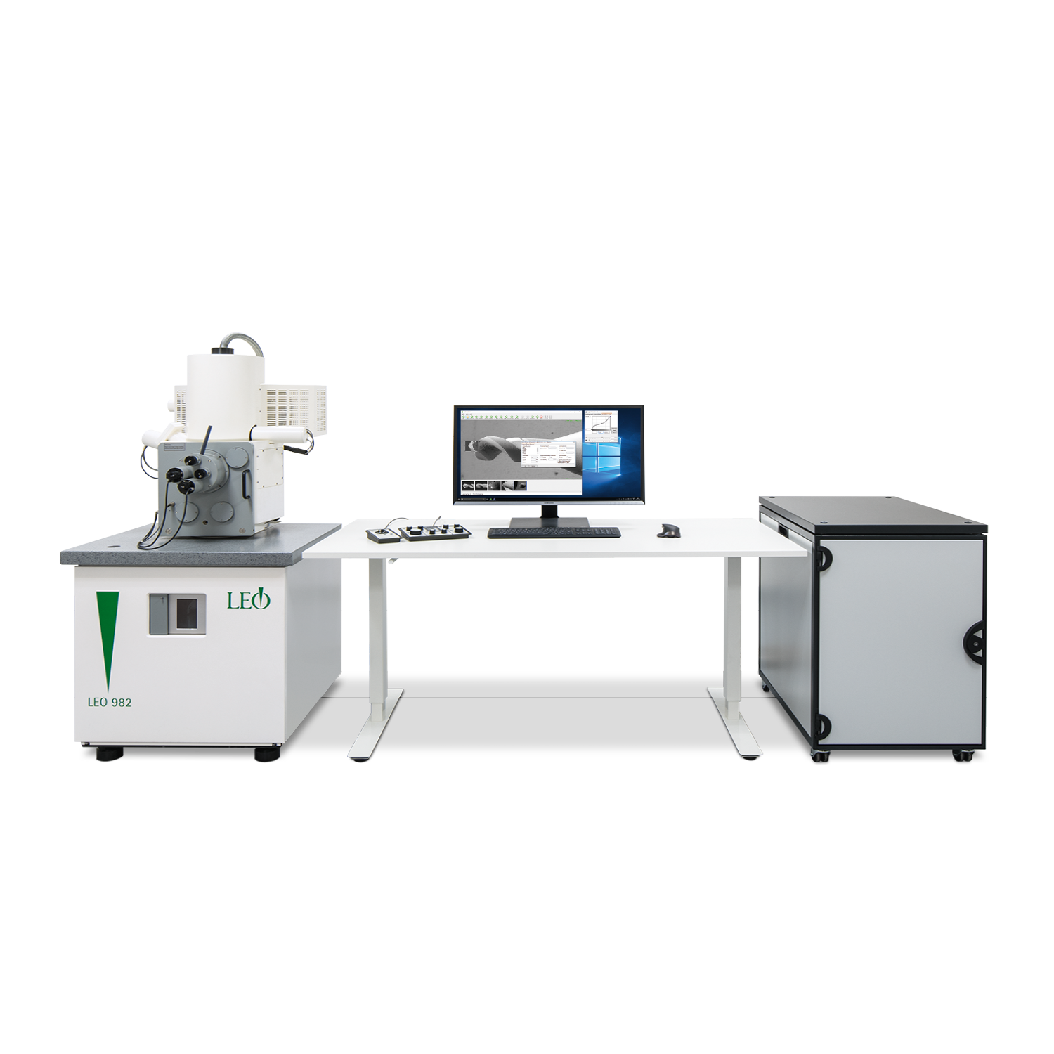
Upgraded microscope with new electronics in separate rack
-
Upgraded microscope with new electronics integrated in the plinth

-
Handset with SEM and trackball panel for function control
-
SEM Control software for controlling functions and for displaying and analyzing measurement data

-
-
-