SEM Modernization
Modernize your SEM to the latest technology
Enhance your trusted electron microscope to meet modern standards! Our SEM Modernization exchanges outdated or not longer servicable add-ons for cutting-edge technology - from acquisition to control and software. Each microscope configuration and installation is carefully tailored to each customer's requirements.
Our modernization is possible for many types of scanning electron microscopes (SEM).
The point electronic SEM modernization adds state of the art performance and keeps the electron microscope serviceable for the next 10+ years. So: Don’t scrap – modernize!
Every modernization is a bespoke solution
We equip your SEM with completely new electronics, software and controls so that the microscope will perform even better for many years to come.
Your microscope will not only remain usable for all existing and future detectors, cameras, etc. We also tailor to your required technical options, technologies and applications in the SEM, up to the packaging solution in the plinth or in a separate rack.
Get in touch with us.
User experiences
What do our users and partners say about us? Read the experiences of scientists and engineers who work with a microscope modernized by point electronic. What were the reasons for an SEM update and what has changed compared to before?
-

“We decided to modernize in order to give our SEM a second life, as its performance was still very good but spare part support from the manufacturer has ended. Sustainability and cost efficiency were key factors in our decision.
The modernization process itself worked well and was perfectly organized by the team of point electronic especially with the great effort of Mr. Grauel and Mr. Reeske.”
Jan Gärtner; NaMLab gGmbH/TU Dresden
about the SEM Modernization of their ZEISS LEO1560
-

"After the modernization with the point electronic upgrade, we were able to take amazingly good pictures with the existing detectors (SE / BSE), in a quality that far exceeded that of the original hardware even at the time of purchase 22 years ago. The operation with the two additional panels including trackball and the sophisticated software control of the device is also a lot of fun to work with. Software features such as the false color-coded contrast and brightness settings make operation so much easier."
Ao. Univ-Prof. Mag. Dr. Bernd Minnich, FRMS; Paris Lodron University Salzburg
about the SEM Modernization of a FEI/PHILIPS ESEM XL-30
-

"The LEO 1560 has been with us since the end of 2001 and has served us very well so far. At the beginning of 2023, we received notification from the manufacturer that the device would be discontinued on October 1, 2023. As the LEO 1560 is a workhorse for us, we were looking for a way to operate the device for longer reliably, without the risk of total failure. This was achieved through modernization (replacement of hardware and software) by point electronic."
Bernd Leibold, Institut für Mikroelektronik Stuttgart IMS
about the SEM Modernization of a LEO 1560
-

"Over the past years, we have modernized two scanning electron microscopes (SEM) and a TEM. [...] The old systems could no longer fulfill the increased requirements in the areas of digital imaging, documentation and analysis as well as IT connection. In addition, there will soon be no spare parts for the devices and therefore no repair options. However, the technical design and the measuring principle of the devices still correspond to the current technical standard. [...] For us, the modernization by point electronic is a more cost-effective and resource-saving alternative to a new purchase."
Frank Steiniger, Electron Microscopy Centre at the University Hospital Jena
about the SEM Modernization of a LEO DSM982 and a LEO Gemini 1530
-

"Over the past four years, we have modernized two scanning electron microscopes (SEM) and a TEM. [...] The old systems could no longer fulfil the increased requirements in the areas of digital imaging, documentation and analysis as well as IT connection. In addition, there will soon be no spare parts for the devices and therefore no repair options. However, the technical design and the measuring principle of the devices still correspond to the current technical standard. [...] For us, the modernization by point electronic is a more cost-effective and resource-saving alternative to a new purchase."
Gaby Esser, Institute of Fusion Energy and Nuclear Waste Management (IFN) at the Forschungszentrum Jülich
about the SEM Modernization of a LEO DSM982
Advantages
The effects of our modernization for your SEM:
Performance
increased performance - from sample insertion to final image
Service
a system that is fully serviceable, incl. 10+ years spare part guarantee for point electronic products
Functions
new techniques and capabilities in the SEM
Add-Ons
Keep established workflows and add-ons
Costs
cost efficient, plus costs savings for acquisition, operation and service
How does the SEM Modernization work?
We optimize your microscope exactly to your needs - but that doesn't mean we exchange the whole system. With our SEM Upgrade, we change everything that needs changing and keep what is important for you.
YOU KEEP
Your column incl. HV-PSU and pumps
Your add-ons, according to functionality and your requirements
(such as EDX, EBSD, etc).
Your established/certified workflows
Your laboratory layout and ancillary equipment
WE ADD
State of the art electronics
User friendly software with efficient workflows
Improved performance
Network compatibility
New techniques
Reduced costs
Environmental friendlyness
How do we do the SEM Modernization?
Once we have configured the upgrade to your needs, specifications and conditions in your lab, we arrange an appointment at your premises:
- We handle the deinstallation of your old equipment.
- We do the on-site installation of the SEM modernization for you, in most cases directly in your lab.
- point electronic Modernizations take around 3 days. This keeps downtime to a minimum.
- With our included training you will be fit for our applications within 1 day.
Arrange a demo with us or request a non-binding quote.
Key Features
Short demonstration of the key features and settings for the scan generation and image acquisition.
Modernization of selected models of all microscope manufacturers
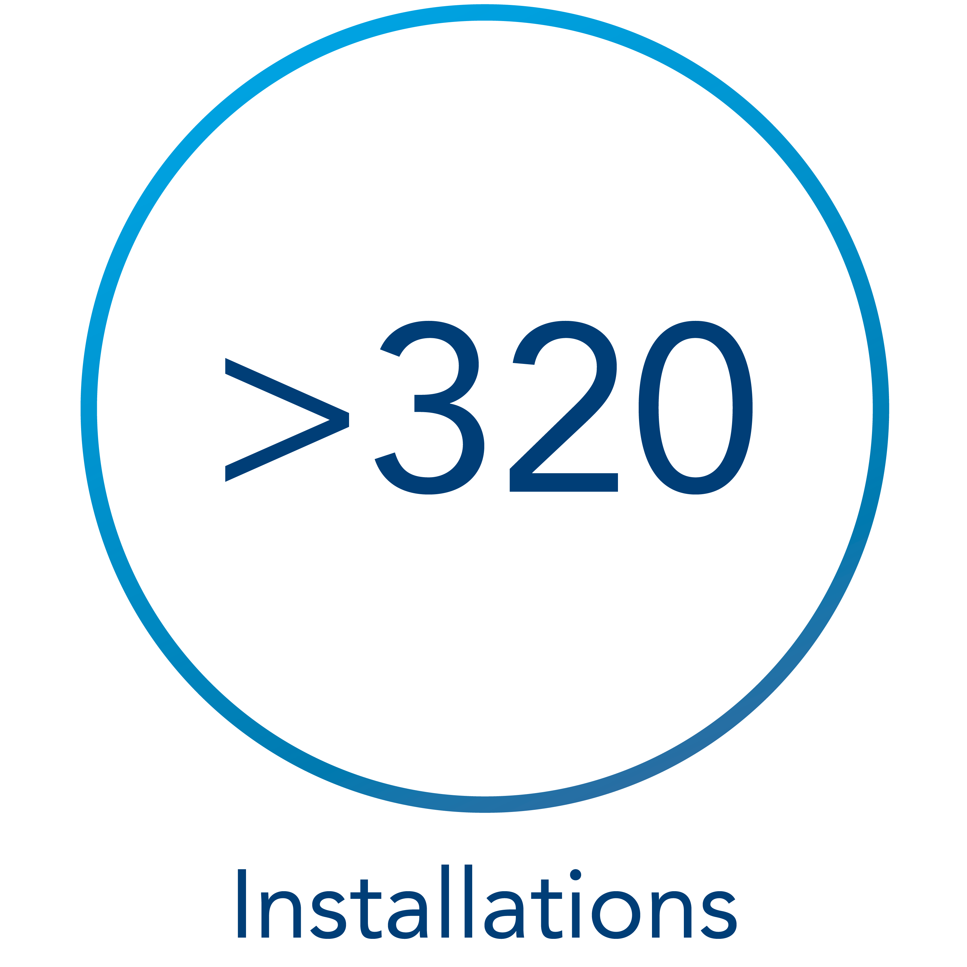
We look back on around 200 installations of our SEM upgrade. For these microscope models we offer SEM modernizations:
Zeiss
- DSM 940, 940A, 960, 962, 982
- LEO / Leica 420, 430, 440
- LEO 1525, 1530, 1530VP, 1550, 1550VP, 1560
- LEO 1430, 1430VP, 1440, 1440VP, 1450, 1450VP, 1455, 1455 VP
- Zeiss SUPRA series with and without VP
- Zeiss ULTRA / ULTRA Plus
- Zeiss Auriga (just SEM column without FIB)
Cambridge
- S90, S120, S200, S250, S260, S360
FEI / TFS
- FEI/Philips XL30, XL30 LaB6, XL30 ESEM, XL30 FEG, XL30 FEG ESEM
- FEI/Philips XL40, XL40 LaB6, XL40 ESEM, XL40 FEG, XL40 FEG ESEM
- FEI/Philips XL20

What is included?
Controls and aquisition
- New SEM control electronics and software
- Support for MicrosoftWindows 7 up to 11, with full network compatibility
- Universal GUI for all upgraded tools independent of manufacturer
- Versatile and fast Scanning system:
- Simultaneous signal acquisition for up to 20 signals (SE, BSE, EDS, WDS, etc.)
- Image resolution of up to 500 MPixels
- Scanning speed down to 10ns dwell time
- Automatic vacuum, gun, stage and detector controls
- New hard panels for column, detector and stage control
- IR chamberscope (for most models)
- PC and display(s) or laptop (optional)
Easy to control - our control panels
We offer two options for the point electronic SEM modernization for control:
You can either combine our SEM Control Panel with our Joystick Panel, or with our Trackball Panel for controlling the stage. Just as it is most practical for you:


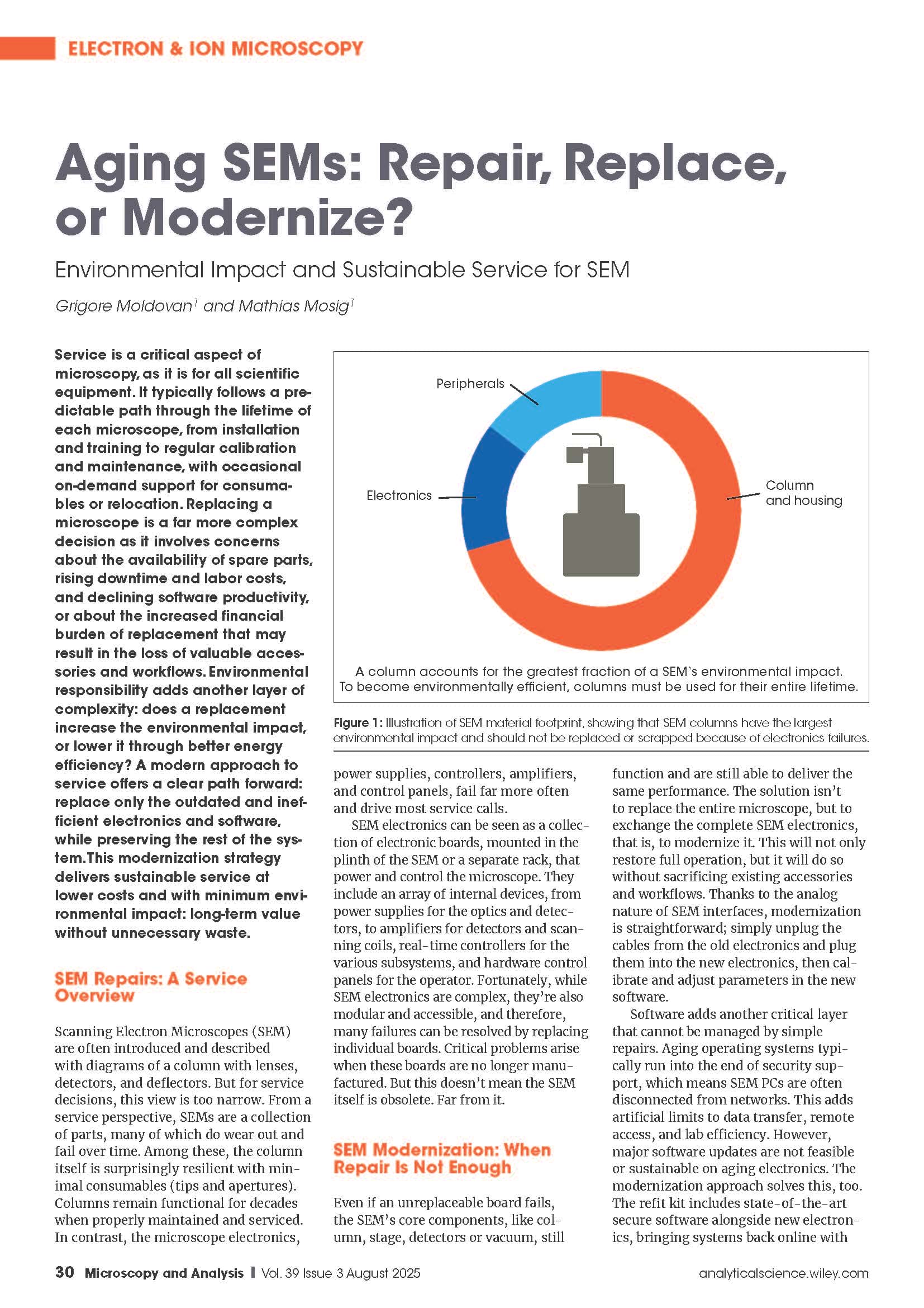
Aging SEMs: Repair, Replace, or Modernize?
Environmental Impact and Sustainable Service for SEM
Article in Microscopy & Analysis, 3/2025, pages 30–32
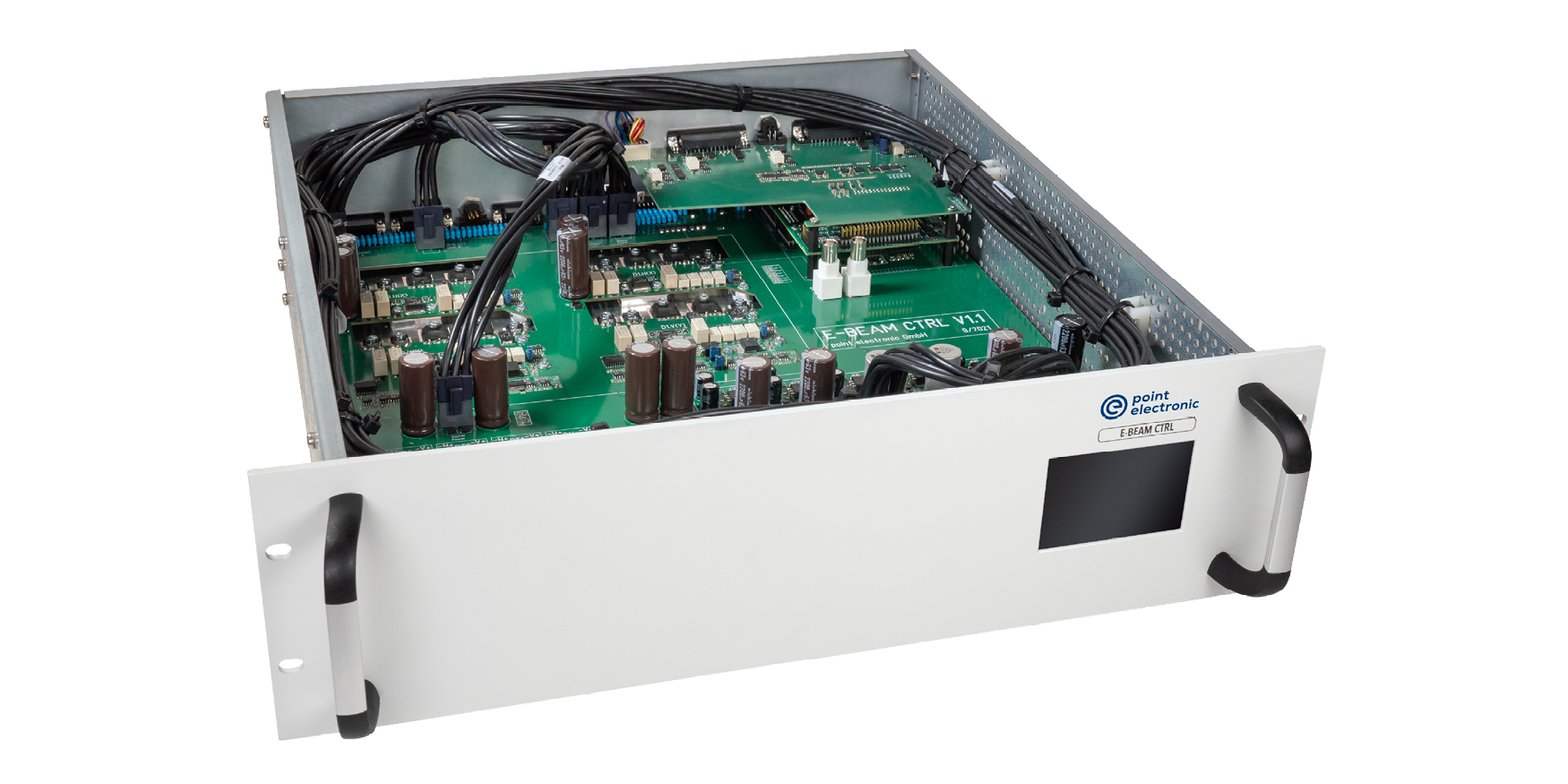
New electronics for the complete SEM
- New control of gun HV and electron detectors
- New bipolar or unipolar high power supply
- New scan module for single or double deflection coils
- 12 digital input signals (X-ray mapping)
- 19 inch, 10U form factor size
- Standard USB 2.0 interface
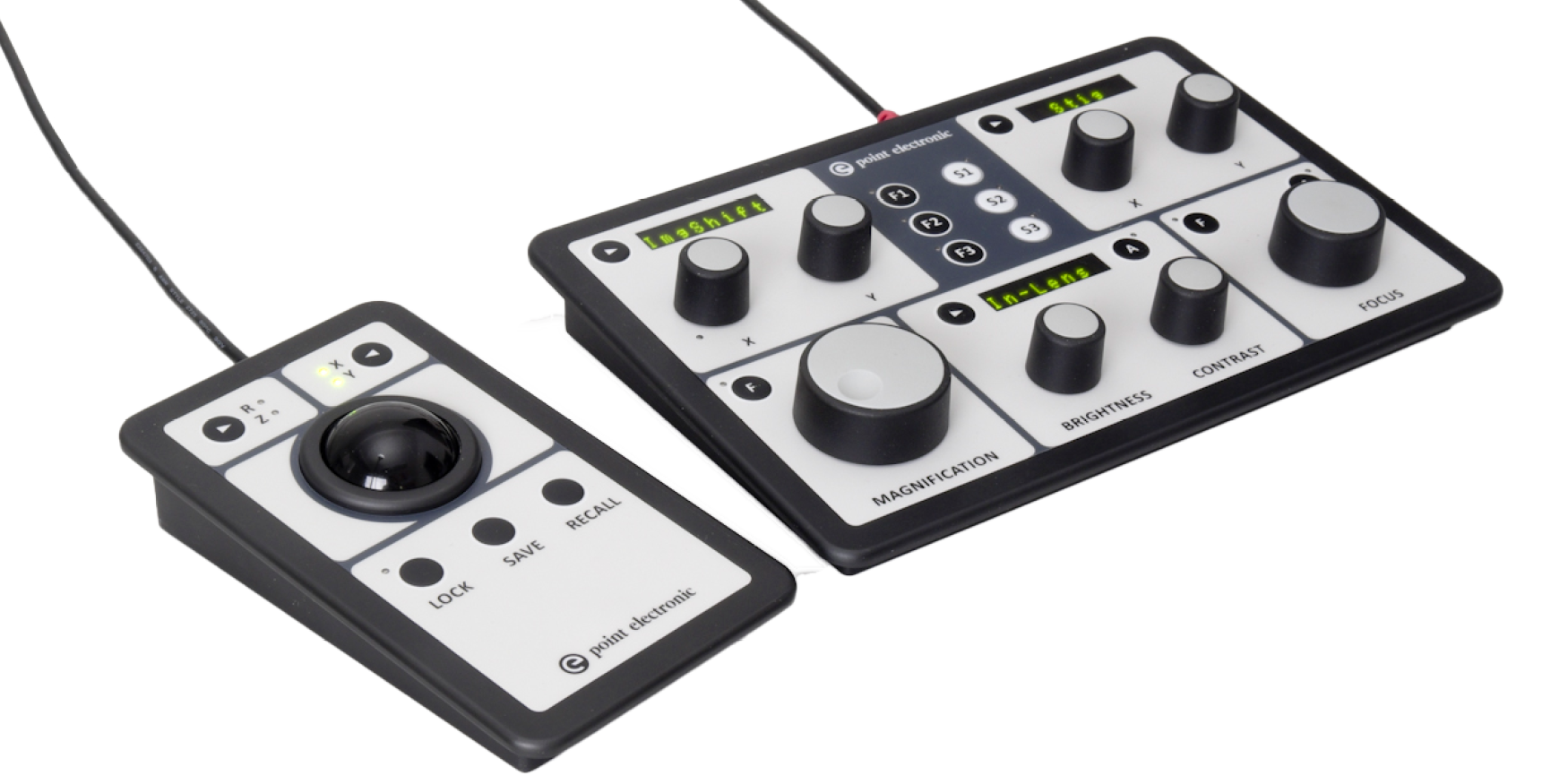
Control panels
- For increased speed and productivity of experienced users
- SEM panel with magnification, focus, image shift, brighness, contrast, stigmatism, etc.
- Stage panel with trackball or joystick, XYRZ locks and store/recall functions
- USB2 controlled and fully integrated wth the microscope control software
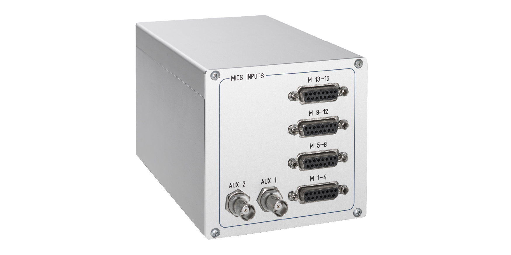
MICS-4 signal amplifier
- For extendend imaging channels, from 4x to 16x
- Channel independendt controls for brightness and contrast
- Advanced input offset and gain controls and calibration
- USB2 controlled and fully integrated with the microscope control software
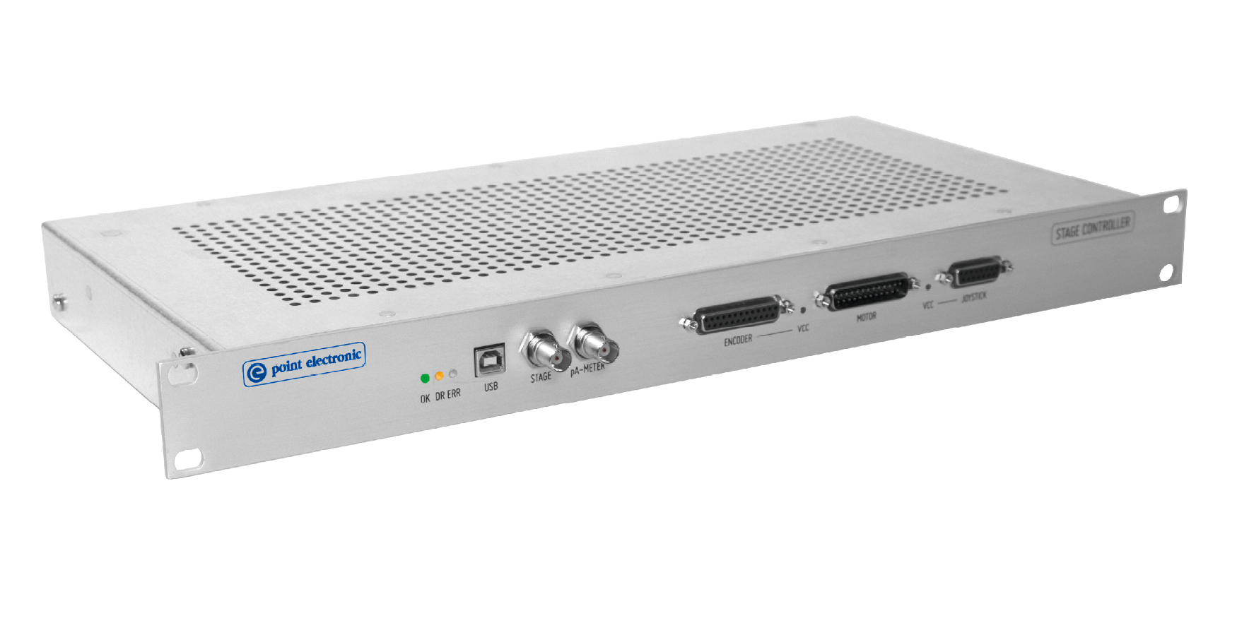
Stage controller
- For automatic positioning and rotation
- Integrated click-and-move, rotation and large area map acquisition
- Configurable software limits for collision avoidance
- USB2 controlled and fully integrated with the microscope control software
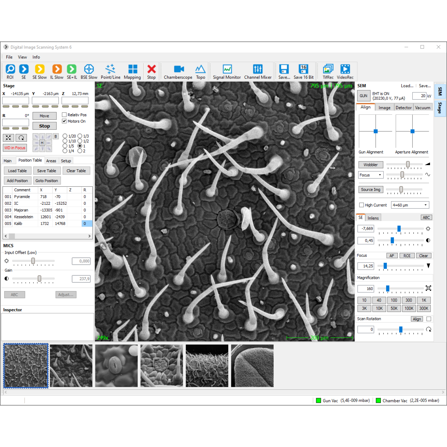
SEM control software
- Control of coils and detectors
- Display of measured values
- Load and save of SEM parameters
- Auto-function for focus/stigmator and brightness/contrast
- TV scan, slow scan, mapping, line scan, point measurements
- Reduced area scan with zoom
- AVI-function with time lapse
- Configurable image acquisition functions for routine actions: Scan buttons can be added, configured and labeled so as signal sources, image resolution, image format
- Integrated vacuum-controller, visualization of vacuum scheme
- Integrated sample stage controller – preset, load and save sample- and stage positions
- Signal monitor with live-histogram
- Display and setting of ir-chamberscope
- Integrated image processing software
- Live 3D reconstruction with BSE-detector (optional)
- AutoSEM for automated capture of multiple sample locations (optional)
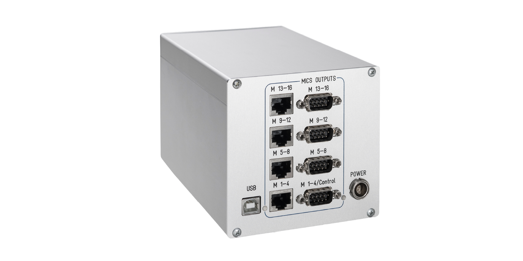
MICS 8 / 12 / 16 signal amplifier
- For extendend imaging channels, by 8x, 12x or 16x
- Channel independendt controls for brightness and contrast
- Advanced input offset and gain controls and calibration
- USB2 controlled and fully integrated with the microscope control software
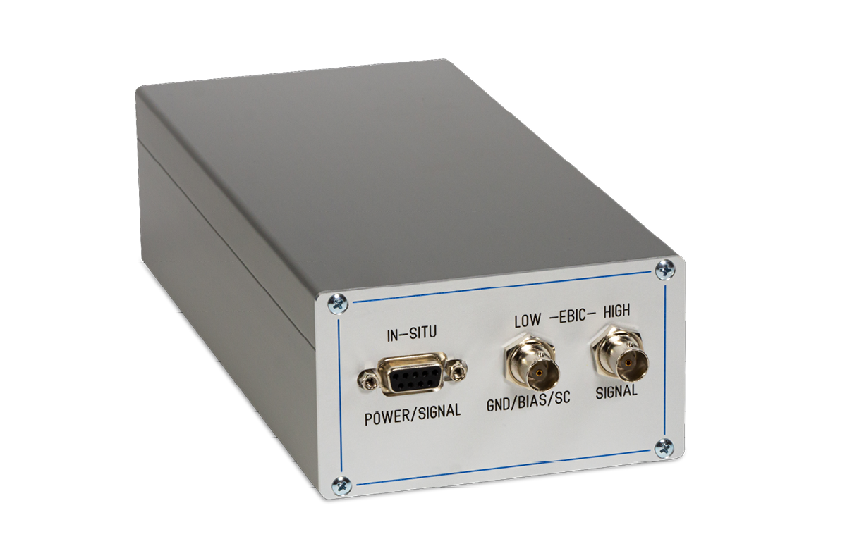
EA amplifiers
- Calibrated high-speed electronics, with in-situ and ex-situ preamplifiers
- Complex multi-stage amplification for imaging, with automatic low-pass filter
- Integrated pico-ammeter for beam current measurements
- Integrated voltage source for biasing
- Integrated current source for compensation
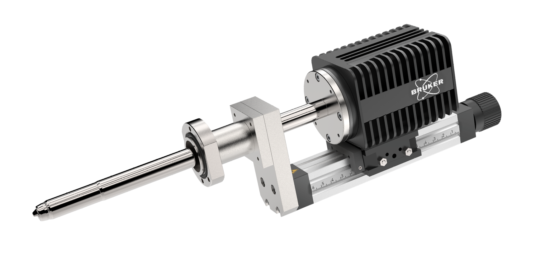
EDS detector
- QUANTAX Compact system by Bruker, containing XFlash® 730M silicon drift detector (SDD), an electronic module, and ESPRIT Compact software
- Allows line scanning and spectral element mapping
- Qualitative and quantitative material analyses
- Fast analysis and reporting
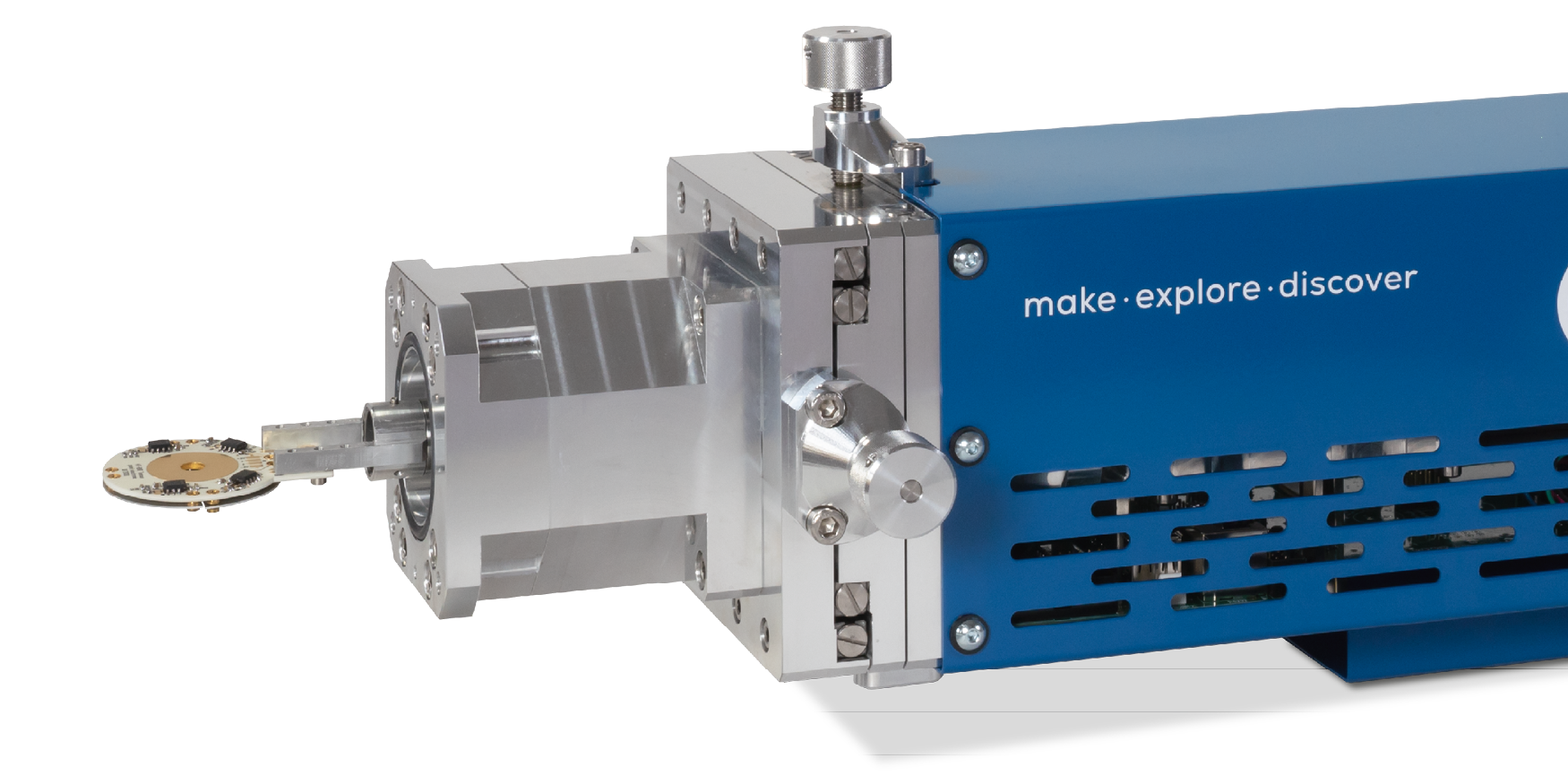
HT BSE detector
- 4Q segmented electrodes with built-in biasing
- Electrodes are light-blind and compatible with laser heating
- Bias voltage to repel secondary electrons and thermal electrons
- Electrodes can be coated in various materials
- easy to disassemble, cleane and recoat
Premium BSE detector
- Four quadrant Si sensor for advanced BSE work
- In-situ preamplification for optimum efficiency and speed
- Optional low-kV sensors
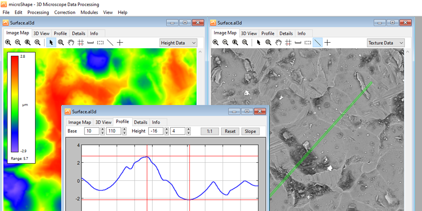
microShape
- Offline 3D viewer of BSE topography data
- Measurement of line profiles, heights, distances and angles in 3D
- 3D data processing including data correction and overlay
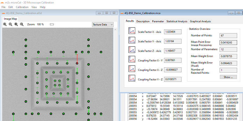
microCal
- Offline 3D calibration and reporting software
- Automated calculation of scales and shearing between all coordinate axes
- Analyses of non-linear scanning deviations
Further options
Additional add-ons and software on demand.
Have a look at our portfolio or contact us.
-
-
-
-
-
-
-
-
-
-
-
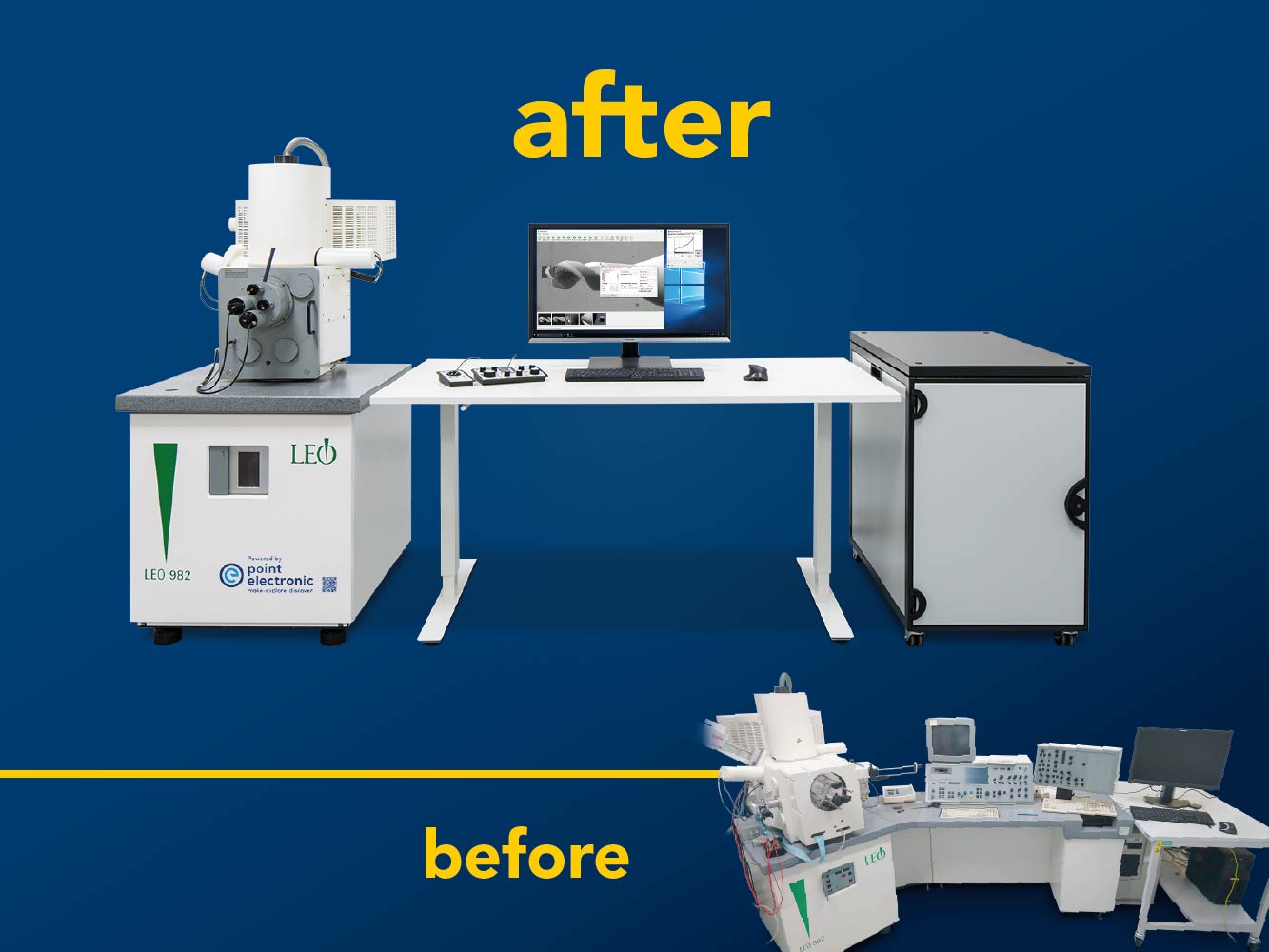
Comparison: SEM before and after the modernization by point electronic GmbH

-
-
Modernized microscope with new electronics in separate rack

-
Modernized microscope with new electronics integrated in the plinth

-
SEM Control software for controlling functions and for displaying and analyzing measurement data

-
SEM Control Panel with Trackball Panel

-
SEM Control Panel with Joystick Panel

-
-